Despite glaucoma’s status as a major cause of blindness, the idea that many individuals may be unaware they have the disease is frightening.
Nearly 50% of glaucoma patients are unaware they have the condition. Reason being, early stages of glaucoma usually do not manifest with symptoms.
Why It’s Critical to Identify Glaucoma Early?
Elevated intraocular pressure is the root cause of glaucoma. The optic nerve is vulnerable to injury from rising intraocular pressure. The issue is that elevated intraocular pressure usually doesn’t cause any noticeable symptoms. Once injury to the optic nerve has started, symptoms typically manifest.
Damage or loss of eyesight can result from glaucoma if not treated properly. Glaucoma causes irreversible eyesight loss. Some people’s vision is already severely impaired when they are diagnosed with glaucoma.
The good news is that if caught early enough, eyesight can be preserved and the disorder can be slowed down. Prevention of eyesight loss is possible with prompt diagnosis and treatment.
Understanding your personal risk factors for glaucoma is crucial. The likelihood of contracting the eye disease varies from person to person. Those at a higher risk of developing glaucoma should visit their eye doctor more often.
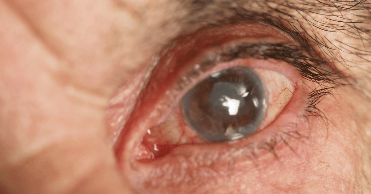
Standard Procedures for Screening for Glaucoma
Diagnosing glaucoma may include a battery of eye tests. In order to make a correct diagnosis, it is necessary to examine the optic nerve thoroughly. Eye specialists are able to choose the best course of treatment for glaucoma with the use of screening instruments.
The gold standard for diagnosing glaucoma is a thorough dilated eye exam. The following visual examinations are part of a complete examination:
Evaluation of Dilated Eye
In order to dilate your pupils, your eye doctor will administer a few drops into each eye during a thorough eye checkup. Your eye doctor can examine the macula, optic nerve, and retina with a slit lamp while your pupils are dilated. Your ophthalmologist will be on the lookout for any changes in the optic nerve’s color or form that could indicate the early stages of glaucoma.
Visual Field Evaluation
To detect changes in vision, a visual field test, also known as a perimeter test, is commonly administered. A visual field test can be conducted in a variety of ways, but all of them serve the same aim. Visual field testing can identify the presence or absence of side vision loss due to glaucoma.
The use of a computerized visual field test is prevalent among these procedures. To check, you must stare directly into a computer screen and mark the spot where a moving light enters your peripheral vision. You might need to have this exam done more than once a year to track any changes in your eyesight.
Tonometry
Another useful diagnostic tool for glaucoma is tonometry, a measurement of intraocular pressure. Your ophthalmologist will use eye drops to numb you while the exam is underway. The eyeballs are gently pressed with a little tool or a puff of air. After that, your ophthalmologist will insert a device into your eye to gauge the intraocular pressure. When checking a patient’s ocular pressure, doctors use the millimeter-Hg (mm-Hg) unit. The typical range for intraocular pressure is 12–22 mm Hg. A reading of 22 mm Hg or higher indicates elevated intraocular pressure.
The majority of glaucoma patients have eye pressures that are greater than 20 mm Hg. There is not a specific level of elevated eye pressure that definitely leads to glaucoma; conversely, there is no lower level of IOP (intraocular eye pressure) that will absolutely eliminate a person’s risk of developing glaucoma. We say that a patient has ocular hypertension (high eye pressure) when their intraocular pressure (IOP) is higher than normal but no other tests rule out glaucoma.
Pachymetry
An additional component of a thorough eye examination may include pachymetry, which evaluates the cornea’s center thickness. Following the application of eye drops, your ophthalmologist will measure the thickness of your cornea using a little device known as a pachymeter. A central cornea that is thicker might affect eye pressure. Pachymetry is a painless and fast procedure.
Inspection of the Drainage Angle
Sometimes, elevated intraocular pressure (IOP) results from improper drainage of intraocular fluid, a condition known as glaucoma. In order to check for glaucoma thoroughly, a gonioscopy may be necessary. During this diagnostic procedure, your eye doctor can see the drainage angle and assess whether it is obstructed. While your doctor examines the space between your cornea and iris, he or she will numb your eyes and put a portable contact lens into each of your eyes.
Also Read: Laser Eye Surgery: A New Perspective on Ophthalmic Use
Analysis of the Fiber Layers of the Optic Nerve Head and the Retinal Nerve
Examining the optic nerve fiber layer (RNFL) and the optic nerve head (ONH), two critical structures located in the back of the eye required for normal vision, is crucial for comprehending the development of glaucoma in patients. Your ophthalmologist can identify changes in ONH and RNFL thickness with the use of a specialized scanning laser. Taking these pictures at regular intervals will give your ophthalmologist a better idea of how fast, how far, and what kind of disease you’re dealing with. Your eye doctor can use this information to better manage your glaucoma.
Why is an examination for Glaucoma even needed?
It may appear daunting to undergo so many tests just to be screened for glaucoma. It’s not unusual. Regardless, you should get all of them done. Diagnosing glaucoma requires more than just measuring ocular pressure. Although elevated intraocular pressure (IOP) is associated with an increased risk of glaucoma, it is not the sole cause of the illness. Your eye doctor will be better able to assess your present state and recommend a treatment plan after doing a thorough eye exam that covers all aspects of your eye health.
Who is a Candidate for Glaucoma Examination?
A correct diagnosis and a comprehensive eye exam are of the utmost importance when it concerns your eyesight.
Diagnosed with glaucoma. What Should You Do Now?
If you are diagnosed with glaucoma, it is crucial to begin treatment promptly in order to reduce the disease’s advancement and preserve your vision.
Conclusion
A baseline comprehensive eye exam should be scheduled for individuals by the age of 40. Your eye doctor may advise glaucoma screening sooner if they consider your eye health and any risk factors.
Please contact our office at +91 81040 35610 to make an appointment with one of our eye specialists at Deevine Eye Care & Multispeciality Centre, an eye hospital in Mumbai, if you have any inquiries regarding the early identification or diagnosis of glaucoma or if it is time for your annual complete eye exam. As an established eye specialist in Ghatkopar, our team at Deevine Eye Care & Multispeciality Centre is dedicated to providing comprehensive eye care services tailored to your needs.